

FOLLOW US
FOLLOW US
FOLLOW US
FOLLOW US
| Download Brochure |
 |
| Navigation Bar |
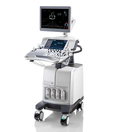
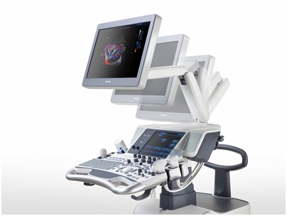
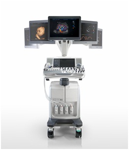
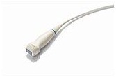
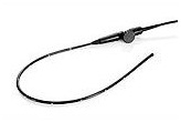
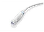
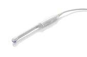
Ultrasound System
From Mind to Heart
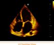
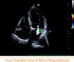
Mindray’s top-of-the-range ultrasound system, DC-8 integrates the best of technology, quality and ergonomics, providing you with a unique scanning experience. Equipped with revolutionary cardiac technology, DC-8 is a result of some of the best minds at work, providing us with answers close to our hearts. With a crystal clear image quality, comprehensive analysis package and a highly user-friendly interface, the DC-8 is your standalone solution for all cardiac examinations.
Performance
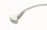
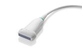
3T Transducer Technology
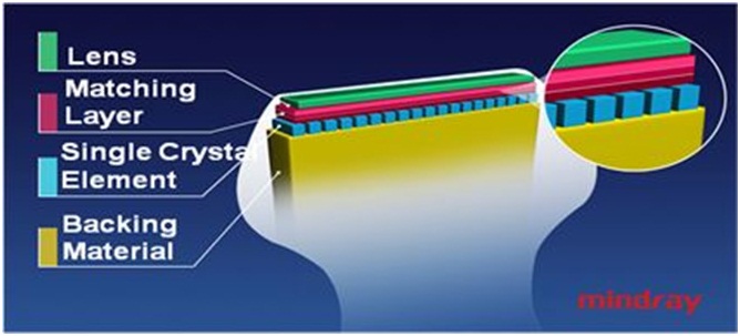
Mindray’s patent transducer technology to increase image bandwidth and transmission efficiency
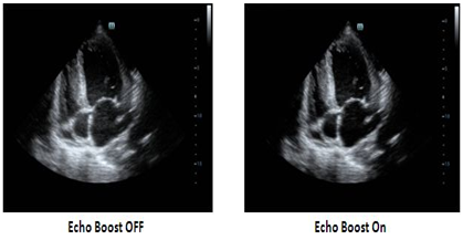
Echo Boost™
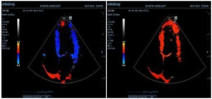
Mindray’s unique adaptive signal processing technology with intelligent echo detection, designed to utilize the native signal-to-noise information to enhance the weak echo signals while suppressing the surrounding clutter noise, providing more balanced image brightness and improved visualization of myocardium tissue layers.
iClear™
Gain improved image quality based on auto structure detection
![]()
with I Clear
![]()
with out I Clear
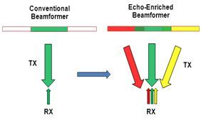
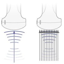
Echo-enriched Beam Forming
Echo-enriched beam former permits the use of traditionally neglected echo signals of adjacent beams to form one finer and stronger imaging beam, providing better ‘out-of-focus’ image resolution and deeper image penetration.
Multi-Beam Formation
Maximum 12 times tasking for one transmitted beam, resulting in excellent time resolution and higher frame rate.
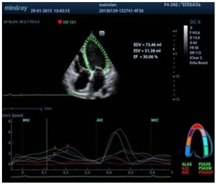
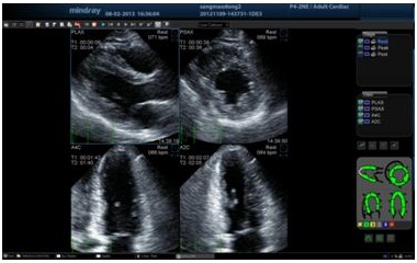
Tissue Tracking with Quantitative Analysis
A simple, quick and non-invasive solution for recognition of left ventricular wall motion abnormalities, TT QA significantly improves the tracking accuracy and effectiveness, significantly controlling the image drift caused by the probe movement and respiration.
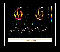
Strain / Strain rate
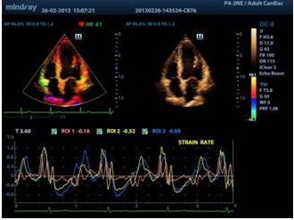
Gain insightful evaluation of ischemic heart disease through strain/ strain rate imaging, providing detailed comparison of different myocardial regions.
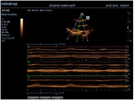
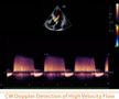
Free Xros M™
Gain precise anatomical observation by freely placing sample lines at any angle. Attain better images through simultaneous display of up to 3 sample lines.
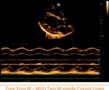
Free Xros CM™
Accurately evaluate myocardial motion at different phases, and simultaneously determine myocardial synchronization. High frame-rate providing you with accurate results
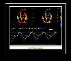
TDI
Tissue Doppler Imaging allows you to quantitatively evaluate local myocardial movement and function, providing complete TDI modes for faster and direct diagnoses.
Stress echo
Gain accurate measurement of the myocardial function in response to external stress or pharmacy
Workflow
Auto LV
Simple measurement procedure for left ventricle enhanced by auto-trace functionality and easy manual correction
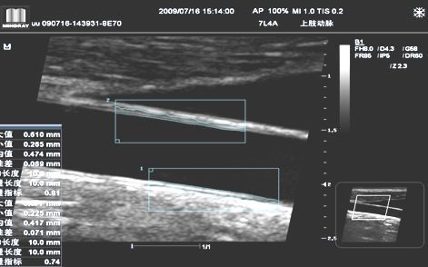
Auto IMT (Intima-Media Thickness)
Auto measurement of anterior and posterior wall thickness providing accurate carotid status
iStorage™
Directly transfer images and reports to PC via network cable as well as convenient measurement & report template edit function
Raw Data
Enables optimum flexibility for post processing of the stored images including parameters adjustment, adding comments and measurements, allowing maximum productivity during scanning
Ergonomics


| Download Brochure |
|
| Navigation Bar |
